Challenge

Cancer cells can become refractory to treatment causing therapy-resistant relapse

Breast cancer is the second leading cause of cancer-related death in women. While early-stage, non-metastatic disease is curable in ~70 to 80 % of patients, advanced breast cancer is considered incurable with currently available therapies.

Treatment is initially effective in 90 % of primary breast cancers and 50 % of metastases. However, only about 10 to 25 % of breast cancer patients achieve complete response after systemic therapy. Even in these cases, if the entire cancer cell population is not eradicated, surviving cancer cells can persist.

The relapsing tumor may remain therapy sensitive and continue to respond to the initial treatment until mechanisms emerge causing stable drug resistance. Residual drug tolerant persister cells cause tumor reoccurrence and therefore are of investigational interest, but are challenging to detect.

Vision

The development of innovative multimodal imaging platforms will contribute to the detection, characterization, and eventual eradication of therapy resistant breast cancer

In REAP we envision to provide imaging systems useful across multiple scales to reveal drug tolerant persister cells in a preclinical setting. We will develop a triple modal two-photon laser scanning – optical coherence – photoacoustic microscopy for in vitro investigations as well as a dual modal optical coherence photoacoustic tomography for in vivo applications. Parallel developments in contrast agents based on biofunctionalized nanoparticles, new lasers, detector technology and real-time data handling aided by deep learning-based analysis will lead to the detection, characterization, and eventual eradication of persister cells.

Therapy resistant breast cancer will be modeled using genetically engineered organoid-based murine mammary tumors.  Newly developed contrast agents will be assayed in vitro and in vivo by the  2-photon laser scanning photoacoustic optical coherence microscopy (2PLS-OC-PAM ) and the optical coherence photoacoustic tomography (OC-PAT) system, respectively, to detect and characterize rare cancer cells.

The triple modal two-photon laser scanning photoacoustic optical coherence (2PLS-OC-PAM) system will provide subcellular and cellular level resolution for up to 2 mm samples. In REAP a triple wavelength laser will be introduced for PAM alongside a new akinetic swept source with both interrogation function for PAM and swept source function for OCT to increase imaging speed. Micro-ring resonator-based (MRR) detector will be developed for superior sensitivity.

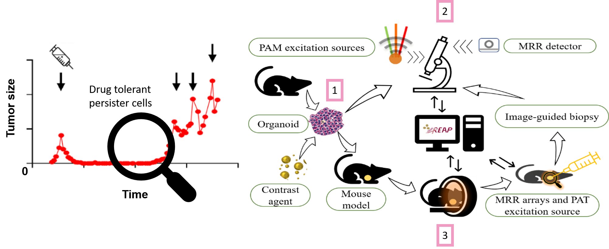
The optical coherence photoacoustic tomography (OC-PAT) system will be used for in vivo studies where increased imaging depth is required. Developments include optical parametric oscillator with high repetition rate and energy for PAT excitation, MRR arrays for real time detection, as well as dual-purpose lasers for all-optical detection photoacoustic interrogation and OCT. Real time image reconstruction algorithms for all the imaging modalities will also be implemented. Moreover, needle tracing algorithm will be implemented for image-guided biopsy. To close the circle, excised biopsies can be further analyzed with the microscopy system (2).

4 Universities

1 Research Institute

4 Industrial Partners

6 European Countries

5 Advisory Board members

Multidisciplinary Fields, 1 Vision

Latest News

Feb. 8th 2024
The REAP M36 Consortium Meeting was hosted in Torino by Prof. Kristen M. Meiburger
on 18 & 19 Jan. Extensive and intriguing discussions on all the
work packages led to clear action plans. The Advisory Board members who
joined virtually gave us comprehensive evaluations and valuable
suggestions on project execution. The Italian hospitality extended from
savory cuisines to a treasure hunt in the city center, where
participants needed to show their dancing skills in front the Egyptian
Museum. The whole consortium is now fully prepared for the upcoming
periodic review, and a fruitful year with many key deliverables.

Contact

Connect with us